| 產品編號 | bsm-54369R |
| 英文名稱 | GEF H1 Recombinant Rabbit mAb |
| 中文名稱 | Rho鳥苷酸交換因子2重組兔單抗 |
| 別 名 | Lbcl1; Lfc; ARHG2; ARHG2_HUMAN; ARHGEF 2; ARHGEF-2; ARHGEF2; GEF; GEF H1; GEF-H1; GEFH1; Guanine nucleotide exchange factor H1; LFP40; Microtubule-regulated Rho-GEF; P40 antibody Proliferating cell nucleolar antigen p40; Protein GEF-H1; Rho guanine nucleotide exchange factor 2; rho/rac guanine nucleotide exchange factor(GEF) 2; rho/rac guanine nucleotide exchange factor 2; rho/rac guanine nucleotide exchange factor. |
| 研究領域 | 腫瘤 免疫學 信號轉導 轉錄調節因子 |
| 抗體來源 | Rabbit |
| 克隆類型 | Recombinant |
| 克 隆 號 | 9D4 |
| 交叉反應 | Human,Mouse,Rat |
| 產品應用 | WB=1:500,IHC-P=1:50-200,IHC-F=1:50-200,IF=1:50-200,ICC/IF=1:50
not yet tested in other applications. optimal dilutions/concentrations should be determined by the end user. |
| 理論分子量 | 112 kDa |
| 檢測分子量 | |
| 細胞定位 | 細胞漿 細胞膜 |
| 性 狀 | Liquid |
| 濃 度 | 1mg/ml |
| 免 疫 原 | KLH conjugated synthetic peptide derived from human GEF H1 |
| 亞 型 | IgG |
| 純化方法 | affinity purified by Protein A |
| 緩 沖 液 | 0.01M TBS (pH7.4) with 1% BSA, 0.02% Proclin300 and 50% Glycerol. |
| 保存條件 | Shipped at 4℃. Store at -20℃ for one year. Avoid repeated freeze/thaw cycles. |
| 注意事項 | This product as supplied is intended for research use only, not for use in human, therapeutic or diagnostic applications. |
| PubMed | PubMed |
| 產品介紹 |
Activates Rho-GTPases by promoting the exchange of GDP for GTP. May be involved in epithelial barrier permeability, cell motility and polarization, dendritic spine morphology, antigen presentation, leukemic cell differentiation, cell cycle regulation, and cancer. Binds Rac-GTPases, but does not seem to promote nucleotide exchange activity toward Rac-GTPases, which was uniquely reported in PubMed:9857026. May stimulate instead the cortical activity of Rac. Inactive toward CDC42, TC10, or Ras-GTPases. Forms an intracellular sensing system along with NOD1 for the detection of microbial effectors during cell invasion by pathogens. Required for RHOA and RIP2 dependent NF-kappaB signaling pathways activation upon S.flexneri cell invasion. Involved not only in sensing peptidoglycan (PGN)-derived muropeptides through NOD1 that is independent of its GEF activity, but also in the activation of NF-kappaB by Shigella effector proteins (IpgB2 and OspB) which requires its GEF activity and the activation of RhoA. Function: Activates Rho-GTPases by promoting the exchange of GDP for GTP. May be involved in epithelial barrier permeability, cell motility and polarization, dendritic spine morphology, antigen presentation, leukemic cell differentiation, cell cycle regulation, and cancer. Binds Rac-GTPases, but does not seem to promote nucleotide exchange activity toward Rac-GTPases, which was uniquely reported in PubMed:9857026. May stimulate instead the cortical activity of Rac. Inactive toward CDC42, TC10, or Ras-GTPases. Forms an intracellular sensing system along with NOD1 for the detection of microbial effectors during cell invasion by pathogens. Required for RHOA and RIP2 dependent NF-kappaB signaling pathways activation upon S.flexneri cell invasion. Involved not only in sensing peptidoglycan (PGN)-derived muropeptides through NOD1 that is independent of its GEF activity, but also in the activation of NF-kappaB by Shigella effector proteins (IpgB2 and OspB) which requires its GEF activity and the activation of RhoA. Subunit: Interacts with 14-3-3 zeta; when phosphorylated at Ser-886. Interacts with the kinases PAK4, AURKA and MAPK1. Interacts with RHOA and RAC1. Interacts with NOD1. Interacts (via the N-terminal zinc finger) with CAPN6 (via domain II). Subcellular Location: Cytoplasm. Cell junction, tight junction. Golgi apparatus. Cytoplasm, cytoskeleton, spindle. Cell projection, ruffle membrane. Note=Localizes to the tips of cortical microtubules of the mitotic spindle during cell division, and is further released upon microtubule depolymerization. Recruited into membrane ruffles induced by S.flexneri at tight junctions of polarized epithelial cells. Post-translational modifications: Phosphorylation of Ser-886 by PAK1 induces binding to protein 14-3-3 zeta, promoting its relocation to microtubules and the inhibition of its activity. Phosphorylated by STK6 and CDK1 during mitosis, which negatively regulates its activity. Phosphorylation by MAPK1 or MAPK3 increases nucleotide exchange activity. Phosphorylation by PAK4 releases GEF-H1 from the microtubules. Similarity: Contains 1 DH (DBL-homology) domain. Contains 1 PH domain. Contains 1 phorbol-ester/DAG-type zinc finger. SWISS: Q92974 Gene ID: 9181 Database links: Entrez Gene: 9181 Human Entrez Gene: 16800 Mouse Omim: 607560 Human SwissProt: Q92974 Human SwissProt: Q60875 Mouse Unigene: 516790 Human Unigene: 239329 Mouse Unigene: 482396 Mouse Unigene: 12255 Rat |
| 產品圖片 |
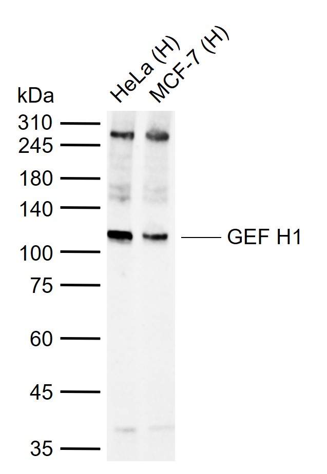
Sample:
Lane 1: Human HeLa cell lysates
Lane 2: Human MCF-7 cell lysates
Primary: Anti-GEF H (bsm-54369R) at 1/500 dilution
Secondary: IRDye800CW Goat Anti-Rabbit IgG at 1/20000 dilution
Predicted band size: 112 kDa
Observed band size: 110 kDa
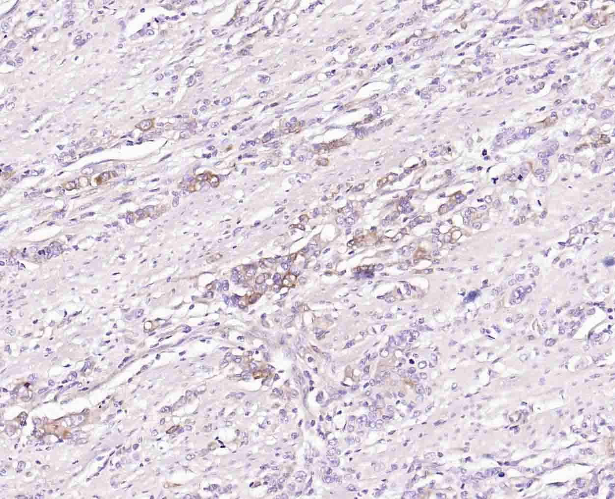
Paraformaldehyde-fixed, paraffin embedded (human gastric carcinoma); Antigen retrieval by boiling in EDTA buffer (Ph9.0) for 15min; Block endogenous peroxidase by 3% hydrogen peroxide for 20 minutes; Blocking buffer (normal goat serum) at 37°C for 30min; Incubation with (GEF H1) Monoclonal Antibody, Unconjugated (bsm-54369R) at 1:50 overnight at 4°C, followed by operating according to SP Kit(Rabbit) (sp-0023) instructionsand DAB staining.

Paraformaldehyde-fixed, paraffin embedded (rat testis); Antigen retrieval by boiling in EDTA buffer (Ph9.0) for 15min; Block endogenous peroxidase by 3% hydrogen peroxide for 20 minutes; Blocking buffer (normal goat serum) at 37°C for 30min; Incubation with (GEF H1) Monoclonal Antibody, Unconjugated (bsm-54369R) at 1:50 overnight at 4°C, followed by operating according to SP Kit(Rabbit) (sp-0023) instructionsand DAB staining.
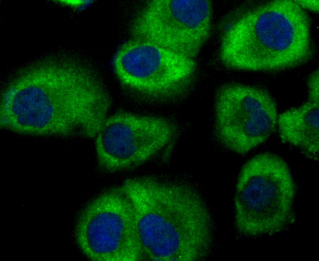
HUVEC cell; 4% Paraformaldehyde-fixed; Triton X-100 at room temperature for 20 min; Blocking buffer (normal goat serum, C-0005) at 37°C for 20 min; Antibody incubation with (GEF H1) monoclonal Antibody, Unconjugated (bsm-54369R) 1:50, 90 minutes at 37°C; followed by a conjugated Goat Anti-Rabbit IgG antibody at 37°C for 90 minutes, DAPI (blue, C02-04002) was used to stain the cell nuclei.
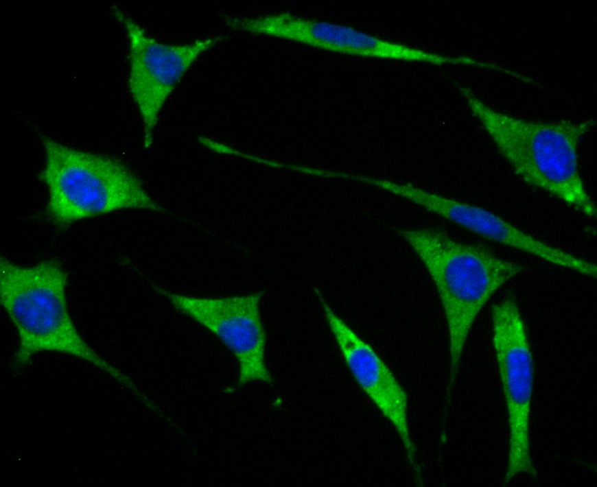
SH-SY5Y cell; 4% Paraformaldehyde-fixed; Triton X-100 at room temperature for 20 min; Blocking buffer (normal goat serum, C-0005) at 37°C for 20 min; Antibody incubation with (GEF H1) monoclonal Antibody, Unconjugated (bsm-54369R) 1:50, 90 minutes at 37°C; followed by a conjugated Goat Anti-Rabbit IgG antibody at 37°C for 90 minutes, DAPI (blue, C02-04002) was used to stain the cell nuclei.
|
| 1、抗體溶解方法 | |
| 2、抗體修復方式 | |
| 3、常用試劑的配制 | |
| 4、免疫組化操作步驟 | |
| 5、免疫組化問題解答 | |
| 6、Western Blotting 操作步驟 | |
| 7、Western Blotting 問題解答 | |
| 8、關于肽鏈的設計 | |
| 9、多肽的溶解與保存 | |
| 10、酶標抗體效價測定程序 | |